Growth Plate Injuries


Growth Plate Injuries
Fractures are a common injury. In fact, there are approximately 6.3 million fractures each year in the USA. There are a variety of types of fractures: compound, tranverse, comminuted, and oblique to name a few. However, fractures involving the growth plates of children are classified differently. Mr Jorge Muniz (physician assistant and comic illustrator) depicts the five types of Salter-Harris fractures (medcomic.com).
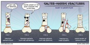
Let’s take a look at each of the five fracture types. I have found through both my own education and my academic career, seeing the relationship of concepts enhances the understanding. Let’s use the mnemonic S – A – L – T – R.
S = separated
A = above
L = lower
T = through
R = rammed
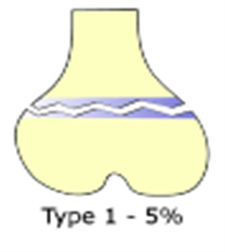
A type I fracture only occurs about 5% of the time. Type 1 corresponds to the letter “S” for “separated.” The growth plate is wider than normal. Obviously a radiograph of both limbs would be needed to know what is “normal.”
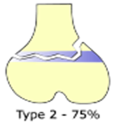
A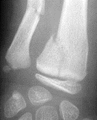 type II fracture is the most common of growth plate injuries. This type occurs about 75% of the time. Type 2 corresponds to the letter “A” for “above” the epiphyseal plate. So the epiphyseal injury goes through the growth plate and above into the metaphysis.
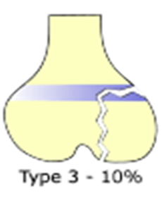
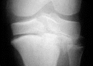
 A type III fracture is less common, only occurring in about 10% of cases. This type corresponds to the letter “L” for “lower.” A type III fracture goes through the growth plate as well as the epiphysis. This type of injury must be reduced to restore smooth articular surface. It most often occurs at the knee or distal tibia.
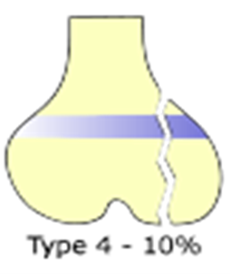
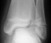
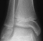
 A type IV fracture is also not very common, only occurring in about 10% of cases. When it does occur, it is most often at the lateral humeral condyle or medial malleolus. This type corresponds to the letter “T” for “through.” “Through” the growth plate as well as into the metaphysis above and the epiphysis below. All three aspects are involved.
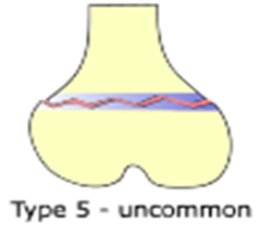
 A type V fracture is very uncommon. This type corresponds to the letter “R” for “rammed.” A type V fracture involves the crushing of the growth plate. Just like a type I fracture, a type V needs a comparative radiograph of the uninvolved side to make the comparison. This injury may appear normal at the time of injury but shows up as growth arrests later.
In summary, clinical signs of a growth plate injury include pain, edema, motion limitations, and sometimes an osseous deformity. Radiographs confirm the presence of epiphyseal injuries but sometimes CT scans, MRIs, or bone scans are needed for more tissue detail. The prognosis of a growth plate injury depends on the area injured, the severity of the injury, and the skeletal age of the patient. Injuries that occur closer to the time of growth plate closure have the best prognosis.
For more cutting-edge orthopedic information on iOrtho+ PREMIUM Mobile App, please visit the learning modules at https://iortho.xyz/
If you would like to learn more about the Mobil–Aider Arthrometer to quantify joint mobility, please visit: https://mobil-aider.com/