Can You Identify Subtle Motor Deficits?


Can You Identify Subtle Motor Deficits?
Anyone who has gone through medical training has thought s/he has had at least one of the diseases studied. You learn the signs and symptoms and suddenly you realize you have some or all of them. In most cases, it is a “false alarm” but do you know how to identify subtle motor deficits that could be an indication of a brain tumor?
A study by Maranhao, Maranhao-Filho, Lima, and Vincent (2010) studied 13 tests for their ability to detect subtle motor deficits to identify possible unilateral/monohemispheric brain tumors. The results of this study were very interested and provide simple tests with respectable metrics for simple screening of a potentially serious concern.
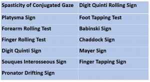
The 13 tests are as follows:
The study was performed in Rio de Janeiro, Brazil. Sixty people with mono-hemispheric cerebral tumors were identified. The control group was 30 people with complaints of vertigo, migraines, or seizures but no tumors. The tests were administered by a physical therapist who was blinded to clinical and imaging data. The descriptions for the performance of the clinical tests were as follows:
| Clinical Test | Description of Performance |
| Spasticity of Conjugate Gaze | Clinician pulls patient eyelid & patient is instructed to exert a small amount of resistance |
| Platysma Sign | Patient asked to retract lower jaw & stretch the neck skin |
| Forearm Rolling Test | Patient makes a fist with each hand, holds forearms horizontal in front of the body, & rolls the arms |
| Finger Rolling Test | Patient extends index fingers pointing towards each other (~1 finger length apart) & rolls fingers around each other |
| Digit Quinti Sign | Patient flexes shoulders to 90°, elbows extended, palms down, abduct fingers |
| Souques Interosseous Sign | Patient raises both arms to 180° of shoulder flexion |
| Pronator Drifting Test | Patient flexes shoulders to 90°, elbows & wrists extended, palms up |
| Mayer Sign | Clinician forcefully flexes patient’s 3rd, 4th, 5th MCP joints. |
| Finger Tapping Sign | Patient taps index finger to distal IP of thumb quickly for 10 seconds |
| Digit Quinti Rolling Sign | Patient extends 5th digits pointing towards each other while other fingers are flexed. 5th digits roll around each other |
| Foot Tapping Test | Patient sits with knee & ankle flexed to 90° & heel on the floor. Patient taps the foot for 10 seconds |
| Babinski Sign | Clinician sweeps lateral border of plantar surface of the foot (heel to digits 4 & 5) with a blunt object |
| Chaddock Sign | Clinician stimulates the dorsolateral aspect of the foot (just inferior & anterior to the lateral malleolus) along the lateral edge of the foot |
The chart below describes the criteria of a positive test and the associated sensitivity (screening metric), specificity (diagnostic metric), and positive/negative predictive values (PV).
| Clinical Test | Criteria for a Positive Test | Sensitivity | Specificity | (+) PV | (-) PV |
| Spasticity of Conjugate Gaze | Eyes deviated to the opposite side of lesion | 0.01 | 1 | 1 | 1 |
| Platysma Sign | Platysma muscle asymmetry | 0.01 | 1 | 1 | 1 |
| Forearm Rolling Test | 1 arm orbits around the other | 0.16 | 1 | 1 | 0.37 |
| Finger Rolling Test | 1 finger orbits around the other | 0.41 | 0.93 | 0.92 | 0.44 |
| Digit Quinti Sign | 5th digit adducts on involved side | 0.51 | 0.70 | 0.77 | 0.42 |
| Souques Interosseous Sign | Involved fingers extend and abduct | 0.23 | 0.80 | 0.70 | 0.34 |
| Pronator Drifting Test | Inability to maintain position for 20-30 seconds. Pronation or downward drift of arm | 0.41 | 0.96 | 0.96 | 0.45 |
| Mayer Sign | Lack of thumb opposition combined with MCP flexion & IP extension | 0.06 | 0.86 | 0.50 | 0.31 |
| Finger Tapping Sign | .5 repetition difference between hands | 0.18 | 0.90 | 0.78 | 0.35 |
| Digit Quinti Rolling Sign | Involved finger orbits less around the other | 0.06 | 0.95 | 0.66 | 0.42 |
| Foot Tapping Test | >5 repetition difference between feet | 0.23 | 0.93 | 0.87 | 0.37 |
| Babinski Sign | Extension of great toe | 0.08 | 1 | 1 | 0.35 |
| Chaddock Sign | Extension of great toe & foot flares | 0.03 | 1 | 1 | 0.34 |
Maranhao et al (2010) sought to identify the most useful tests for detection of brain tumors. Despite positive results for the Spasticity of Conjugate Gaze, Platysma Sign, Babinski Sign, Chaddock Sign, and Mayer Sign, they all are indicative of pyramidal tract dysfunction and had low indices for monohemispheric brain tumors. The Digit Quinti Sign, Pronator Drifting Test, and Finger Rolling Test was deemed the most sensitive tests. While the Digit Quinti Sign, Pronator Drifting Test, and Finger Rolling Test had the greatest specificity. These data are consistent with the research of Alter (1973), Sawyer et al (1993), Anderson et al (2005), and Yamamoto (1995). Clinicians should be aware of these tests to provide simple methods to detect subtle motor deficits that may warrant neuroimaging.
For more cutting edge orthopedic information, subscribe to iOrtho+ Premium Mobile Web App, at https://iortho.xyz/
- Alter M. The Quinti digiti sign of mild hemiparesis. 1973;23:503-505
- American Cancer Society. https://www.cancer.org/research/cancer-facts-statistics/all-cancer-facts-figures/cancer-facts-figures-2020.html
- Anderson NE, Mason DF, Fink JN, Bergin PS, Charleston AJ, Gamble GD. Detection of focal cerebral hemisphere lesions using the neurological examination. Journal of Neurology Neurosurgery & Psychiatry. 2005;76:545-549.
- Goodman, C, Snyder, T. Differential Diagnosis in Physical Therapy, WB Saunders Co, Phila, 3rd ed, 2000
- Gulick DT. iOrtho+ Mobile App. DTG Enterprises LLC. 2020
- Gulick, DT. OrthoNotes, 4th FA Davis Publishing, Philadelphia. 2018
- Maranhao ET, Maranhao-Filho P, Lima MA, Vincent MB. Can Clinical Tests Detect Early Signs of Monohemispheric Brain Tumors? Journal of Neurologic Physical Therapy. 2010;34(3):145-149
- Sawyer RN, Hanna JP, Ruff RL, Leigh RJ. Asymmetry of forearm rolling as a sign of unilateral cerebral dysfunction. Neurology. 1993;43:1596-1598
- Walsh Flanagan K & Cuppett M. Medical Conditions in the Athlete. 3rd ed, Human Kinetics, Champaign IL. 2017
- Yamamoto T. Forearm rolling test. Neurology. 1995;45:2299