Anatomy is the Root of Differential Diagnosis


Anatomy is the Root of Differential Diagnosis
As clinicians we frequently hear about how we “create“ pain in our examination or treatment sessions. In the examination process “reproduction” of pain….the pain that brought them to seek care…. is very important. The “reproduction” of their symptoms guides our differential diagnosis.
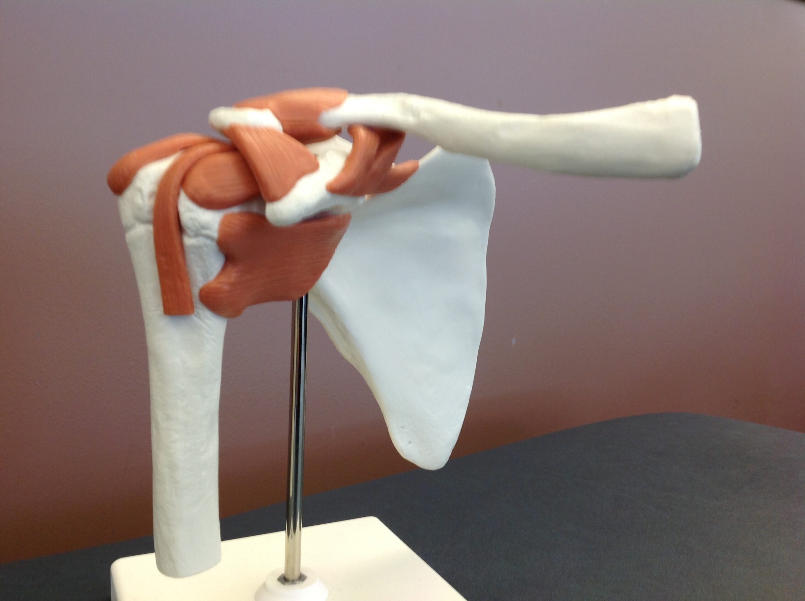
Anatomy is the root of this process. Knowing the attachment sites and actions are critical. We must know the attachment sites to effectively perform surface palpation of the muscles. Once we are able to reproduces the pain, the next step is to ask “can you put one finger on the place where you just experienced pain?” This blog will focus on the isolation and differentiation of muscle actions. To demonstrate the process, we will use motions in the cardinal planes of the shoulder.
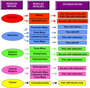
In the sagittal plane we have flexion and extension. Muscles involved in flexion include coracobrachialis, pectoralis major, biceps, and anterior deltoid. Of these muscles, the coracobrachialis is the only pure shoulder flexor. The pectoralis major is also a horizontal adductor and internal rotator. Whereas the biceps is an assistant mover at the shoulder and a prime mover at the elbow. So placing the elbow in maximal flexion and the shoulder in 90 degrees of flexion will put the biceps on slack. The anterior deltoid also contributes to horizontal adduction. So testing horizontal adduction will isolate the anterior deltoid from the other muscles. Finally, the supraspinatus initiates flexion but its alignment in the plane of the scapula does not give it a good mechanical advantage in the sagittal plane.
Shoulder extension involves triceps, posterior deltoid, latissimus, and teres major. To isolate these muscles, the posterior deltoid can be incriminated via supported horizontal abduction with the elbow extended. This position puts the triceps in an active insufficient position (on slack) to minimize its ability to contribute. Testing the triceps via elbow extension will take the deltoid out of the picture. Likewise, testing the latissimus with the elbow extended will relegate the triceps to an assisted mover at the shoulder (biarticulate muscles are always prime movers distally). Together the latissimus and teres major are also internal rotators so resisting internal rotation with the shoulder in neutral will eliminate the triceps and posterior deltoid.
In the coronal plane, we have shoulder abduction and adduction. Abduction is performed via the supraspinatus, deltoid, and may have contributions from the biceps and coracobrachialis with increasing external rotation (influencing the line of pull). To incriminate the various heads of the deltoid, one can support the shoulder in 90 degrees of abduction and resist horizontal adduction for the anterior deltoid and horizontal abduction for the posterior deltoid. Supraspinatus not only initiates abduction but it contributes throughout the range of motion. Thus, to differentiate the supraspinatus verses middle deltoid involvement it is best determined via palpation.
Adduction is performed by the pectoralis major, teres minor, latissimus, and teres major. If the pectoralis major is involved, there may also be pain with flexion and internal rotation. If the teres minor or major are involved, there will be pain with external rotation or internal rotation, respectively. If the latissimus is involved, there will be pain with extension and internal rotation.
In the transverse plane we have shoulder internal (IR) and external rotation (ER). Internal rotators include the pectoralis major, anterior deltoid, latissimus, teres major, and subscapularis. The pectoralis major and anterior deltoid also perform horizontal adduction. While the latissimus and teres major contribute to shoulder extension. Rigsby, Sitler, and Kelly (2010) demonstrated the ability to distinguish involvement of the upper and lower fibers of the subscapularis. When resisting IR with the arm at 90 degrees of elevation, the lower subscapularis fibers are more active. Whereas, when resisting IR at 45 degrees of elevation, the upper fibers are more involved.
Muscles contributing to external rotation are the supraspinatus, infraspinatus, teres minor, and posterior deltoid. Approximately 24% of supraspinatus tears also involve the infraspinatus tendon due to the congruent attachment at the anterosuperior edge of the humerus. Distinguishing the supraspinatus and infraspinatus can be challenging. Clinicians should he aware that if testing ER with the shoulder at 90 degrees of abduction, a drop in the elbow position (inability to maintain abduction) implicates the supraspinatus, whereas the inability to maintain ER may point to the infraspinatus. Furthermore, EMG studies have found higher activation of the infraspinatus in actions involving shoulder flexion. In addition, the speed of the shoulder movement may be another variable to isolate the infraspinatus. The late cocking phase of a baseball throw has a high infraspinatus to supraspinatus ratio. Hughes, Taylor, and Green (2015) suggested faster isotonic motions may help isolate the infraspinatus.
In summary, knowing anatomy and muscle actions are the foundation of clinical practice. Given the majority of muscles contribute to more than one action, being able to differentiate the actions can be valuable in the identification of the pathology. Likewise, positioning of adjacent joints can alter the length-tension relationship of a biarticulate muscle and influence its ability to contribute to an action. Palpation and clustering of clinical tests can further support a given diagnosis. I hope this exercise with the shoulder will help in the application of differentiating pathology in other joints.
For more cutting-edge orthopedic information on iOrtho+ PREMIUM Responsive Web App, please visit prior blog posts and the learning modules at https://iortho.xyz/
If you would like to learn more about the Mobil-Aider orthopedic arthrometer to quantify joint mobility, please visit: https://mobil-aider.com/
References:
- Gulick DT. iOrtho+ Mobile App. DTG Enterprises LLC. 2020
- Gulick, DT. OrthoNotes, 4th FA Davis Publishing, Philadelphia. 2018
- Hughes P, Taylor NF, Green R. Identifying shoulder testing positions and movements that isolate infraspinatus from supraspinatus. Journal of Physical Therapy Reviews. 2015;20(2):73-85
- Rigsby R, Sitler M, Kelly JD. Subscapularis tendon integrity: an examination of shoulder index tests. Journal of Athletic Training. 2010;45(4):404-406