
The Upper Limb Tension Tests (ULTTs) are designed to assess the length and mobility of the neurological structures of upper limb. These tests were first described by Elvey in 1994. The tests include specific positions of the shoulder, elbow, forearm, wrist and fingers to bias each particular nerve. The ULTT’s are equivalent to the various straight leg raise tests for the lower limb.
There are 3 upper limb tension tests: median (ULTT1), radial (ULTT2), and ulnar (ULTT3). Each of the tension tests will be described with the available statistical data.
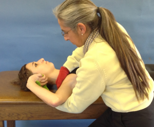
Median nerve (ULTT 1) 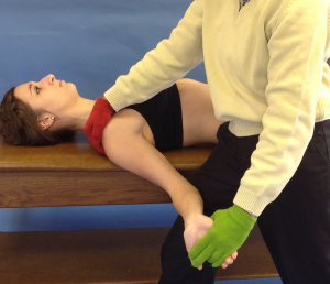
The median nerve originates from the lateral and medial cords of the brachial plexus. The lateral cord has contributions from cervical roots 5, 6, and 7. Whereas the medial cord has contributions from roots C7 and T1. Of interest is the fact that the median nerve is the only nerve to pass through the carpal tunnel.
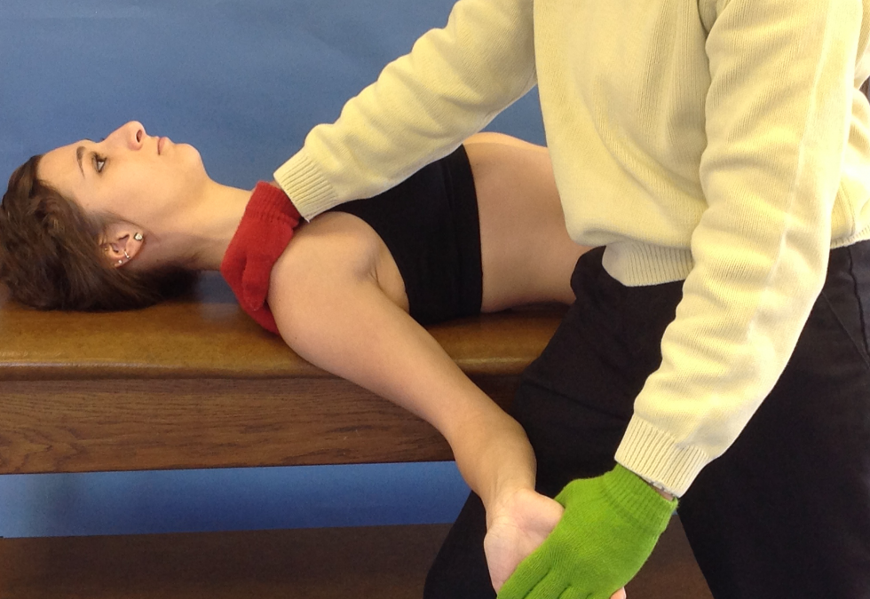
To assess the median nerve, the patient would begin in supine or in sitting. The components are as follows:
- contralateral cervical sidebending
- ipsilateral shoulder depressed
- shoulder abduction in external rotation
- elbow extension with forearm supinated
- wrist/fingers (including thumb) extension
Clinicians have the option to impart these components from proximal to distal or from distal to proximal. Some clinicians opt to implement elbow extension last to use it as a barometer for the presence of a restriction. If elbow extension is added last, the angle at which a limitation is identified can be measured to quantify a restriction.
A positive test for median nerve pathology is pain or paresthesia into median nerve distribution, i.e. first two to three digits of the hand. The data below reveals the high sensitivity and low specificity of the median nerve neural tension test. Thus, the upper limb tension test for the median nerve is very good at ruling out problems with the median nerve. However, it does not have strong diagnostic value. It is also very important to notice the impact of combining various tests to identify cervical spine pathology
- Sensitivity = 94-97%, Specificity = 22%
- (+) Likelihood ratio = 1.3
- (-) Likelihood ratio = .012
- Reliability (Kappa) = 0.76
Combinations: Median nerve, Spurling, Distraction, & Ipsilateral c-spine rotation <60 degrees:
- Any 2 (+) tests: Sensitivity = 39%, Specificity = 56%; (+) Likelihood ratio = 0.88
- Any 3 (+) tests: Sensitivity = 39%, Specificity = 94%; (+) Likelihood ratio = 6.1
- All 4 (+) tests: Sensitivity = 24%, Specificity = 99%; (+) Likelihood ratio = 30.3
Radial nerve (ULTT 2)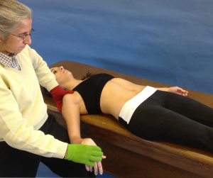
The radial nerve supplies the posterior portion of the upper limb. It innervates the medial and lateral heads of the triceps muscle, as well all 12 muscles in the posterior forearm. The radial nerve is composed of fibers from the ventral roots of spinal nerves C5, C6, C7, C8, and T1.
To assess the radial nerve, the patient could be in supine or sitting. The components are as follows:
- contralateral cervical sidebending
- ipsilateral shoulder is depressed
- arm is extended with elbow extended
- forearm is pronated
- wrist is flexed
- metacarpal phalangeal joints are flexed & the interphalangeal joints are extended.
Again, some clinicians implement these parameters proximal to distal, others distal to proximal, and there is always the option to add elbow extension last to measure the degree of which elbow flexion produces symptoms.
The test is positive with pain or paresthesia into radial nerve distribution of upper extremity, i.e. posterior aspect of the forearm and hand. Similar to the median nerve testing, radial nerve sensitivity is high while specificity is low. Thus, the radial ULTT serves as a good screening tool.
- Sensitivity = 72-97%
- Specificity = 33%
- (+) Likelihood ratio = 1.1
- (-) Likelihood ratio = 0.85
- Reliability (Kappa) = 0.83
 Ulnar nerve (ULTT 3)
The ulnar nerve is an unprotected nerve. Typically, nerves are protected by muscle and/or bone but the ulnar nerve is not, so injury is common. We have all experienced bumping our “funny bone” and having paresthesias into the ring and pinky finger. This is the result of the ulnar nerve being trapped between the skin and the medial epicondyle. The ulnar nerve originates from the C8 and T1 nerve roots (sometime C7 also contributes). The ulnar nerve provides sensory innervation to the 4th and 5th digits, as well as motor branches to innervate at least 11 muscle of the forearm and hand (including FCU, opponens digiti minimi, dorsal interossei, etc).
To assess the ulnar nerve, the patient could be in supine or sitting. The components are as follows:
- ipsilateral shoulder is depressed
- abduct the shoulder to 90° with external rotation
- flex the elbow with the forearm pronated
- extend wrist/fingers in an attempt to place the palm of the hand on the ipsilateral ear.
Another option some clinicians prefer is to stabilize shoulder/scapula and extend the wrist, then use the amount of elbow flexion as the final motion to quantify ulnar nerve tension.
The test is positive with pain or paresthesia into ulnar nerve distribution of upper extremity, i.e. digits 4 and 5.
There are 2 problems with ulnar nerve testing:
- there are no data on sensitivity and specificity
- some individuals do not have enough shoulder mobility to assume the test position
 In this case, Manvell et al (2015) identified an alternative technique. The researchers reported data to demonstrate this technique was effective at incriminating the ulnar nerve and differentiating it from the median and radial nerves. The technique has the patient place the palm of the hand on the side of the rib cage. The position of the arm is:
- abduction with internal rotation
- elbow flexion with forearm pronation
- wrist/finger extension
In spite of the instructions provided in this blog for the clinician, these techniques can also be taught to a patient to perform neural flossing on their own. Neural flossing is not nerve stretching! Flossing is a gentle technique used to glide the peripheral nerve to increase mobility and reduce pain. This technique can be used to perform the specific positions as part of a home exercise program. All of these techniques, complete with videos are available in iOrtho+ Premium Mobile App.
For more cutting edge orthopedic information in iOrtho+ Premium Mobile App, please visit the learning modules at https://iortho.xyz/
- Arguisuelas Martinez MD, Lopez Cubas C, Lluch Girbes E. Ulnar nerve neurodynamic test: Study of the normal sensory response in asymptomatic individuals. JOSPT. 2014;44(6):450-456
- Butler DS: Mobilisation of the nervous system, Melbourne, 1991, Churchill Livingstone.
- Coppieters MW, Stappaerts KH, Everaert DG, Staes FF. Addition of test components during neurodynamic testing: effect of ROM & sensory responses. Journal of Orthopedics & Sports Physical Therapy. 2001;31:226-237
- Coppieters M, Stappaerts K, Janssens K. Reliability & detecting onset of pain & submaximal pain during neural provocation testing of the upper quadrant. Physiologic Research International. 2002;7:34-42
- Elvey RL: The investigation of arm pain. In Boyling JD, Palastanga N (eds): Grieve’s modern manual therapy: the vertebral column, 2nd ed. Edinburgh, 1994, Churchill Livingstone
- Garmer DA, Jones A, McHorse KJ. The ulnar nerve bias upper limb neurodynamic tension test: An investigation of responses in asymptomatic subjects. APTA Conference Abstract, Cincinnati OH 2002
- Gulick DT. iOrtho+ Mobile App. DTG Enterprises LLC. 2020
- Gulick, DT. OrthoNotes, 4th FA Davis Publishing, Philadelphia. 2018
- Kleinrensink GJ, Stoeckart R, Mulder PGH, Hock GVD, Broek TH, Vleeming A, Snijders CJ. Upper limb tension tests as tools in the diagnosis of nerve & plexus lesions: Anatomical & biomechanical aspects. Clinical Biomechanics. 2000;15:9-14.
- Manvell JJ, Manvell N, Snodgrass SJ, Reid SA. Improving the radial nerve neurodynamic test: An observation of tension of the radial, median, & ulnar nerves during upper limb positioning. Manual Therapy. 2015
- McClellan DL, Swash M. Longitudinal sliding of the median nerve during movements of the upper limb. J Neurol Neurosurgery Psychiatry, 1976;39:566-569
- McClellan DL. Longitudinal sliding of the median nerve during hand movements: a contributory factor in entrapment neuropath. Lancet, 1975;15(7907)633-634
- Wainner RS, Fritz JM, Irrgang JJ, et al. Reliability & diagnostic accuracy of the clinical examination & patient self-report measures for cervical radiculopathy. Spine 2003;28:52-62