
Methods of Assessing Wrist Stability


Radiocarpal Joint
The wrist, AKA the radiocarpal joint, is a synovial joint composed of the radius and proximal row of carpal bones (scaphoid, lunate, triquetrum, and pisiform). Movement occurs along two planes to produce flexion, extension, adduction (ulnar deviation), and abduction (radial deviation). There are a variety of possible wrist injuries but the clinician should focus on the most critical possibilities first. Ruling out fracture, dislocation, and ligamentous instability are paramount. Although imaging can be performed to contribute to the assessment, multiple clinical tests are at your disposal.
Approximately 70% of all carpal fractures are of the scaphoid. The mechanism of injury is most often a fall on out-stretching hand (FOOSH) with forceful wrist in extension in radial deviation. A proximal fracture have a worse prognosis than distal fractures because of blood supply to the scaphoid. Plain films frequently do not reveal the fracture so a scaphoid injury should be treated as a fracture until proven otherwise. Injuries to the distal pole occur in about 5-10% of cases and is not typically a problem. A middle pole fracture occurs about 70-80% of the time and surgery may accelerate healing. Whereas, proximal pole fractures occur about 15% of the time and are the highest risk for non-union. The clinical tests used to assess the scaphoid is the clamp sign and the axial loading test.
Scaphoid Fracture
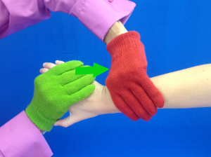 The clamp technique places the forearm in pronation and the wrist in extension. A longitudinal load is applied through a “handshake” like position such that ulnar deviation can be facilitated.
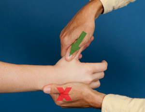
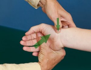
The axial load test is performed with the client sitting and the arm supported on the table. The clinician passively abducts and extensions the metacarpal phalangeal (MCP) joint of the thumb. An axial load is then applied to the 1st carpal metacarpal (CMC) joint. Both tests would be deemed positive if there is pain in the anatomic snuffbox, i.e. location of the scaphoid.
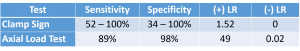 The statistics for clamp sign are highly variable. This is often a problem when a decision on the positivity of a test is based on a subjective assessment of pain. Interestingly the axial load test is statistically better than the traction type maneuver of the clamp sign.
Ligamentous Laxity
The scapholunate (SL) ligament has been referred to as the ACL of the wrist because of its important in wrist stability. Normally, the scaphoid and the lunate bones roll and glide together with the tight ligamentous connection. When a SL ligament tear occurs, the scaphoid bends forward (flexes) and the lunate bends backwards (extends) and a gap may form between the bones. Mechanism of injury for the SL ligament is similar to that of the scaphoid bone, i.e. FOOSH. In fact, fractures frequently accompany SL ligament injuries. Regular radiographs for the SL ligament are frequently normal but a stress film may show a gap between the scaphoid and the lunate. If left untreated, over time the capitate slides between the scaphoid and lunate. This is known as SLAC, scaphoid lunate advanced collapse. Once this ligament is injured it is really tough to get any kind of stability in weight-bearing through the injured hand.
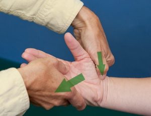 Symptoms of SL ligament injury may include radial side wrist pain, swelling, weak grip, and/or snapping/popping in wrist. Two tests may be used to assess the instability of the scaphoid and lunate. The Watson test can be performed by placing the thumb over the palmar aspect of the distal pole of scaphoid and wrapping the fingers around the distal radius for stabilization. A constant pressure is maintained with the examining thumb as the wrist is moved from a position of extension/ulnar deviation (left) to flexion/radial deviation (right), and back again. This movement pattern is similar to the wrist motions of the PNF diagonal 1. A positive test is dorsal wrist pain or a clunk/shift may indicate instability of the SL ligament. Sensitivity is 69% and specificity is 64-68%. Thus, this test is neither a strong screening tool or diagnostic tool but there is no other dynamic option.
The Murphy sign is used to assess for a lunate dislo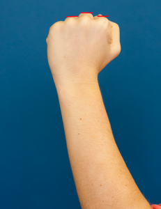 cation which may occur with a SL ligament tear. Under normal circumstances when one views the hand in the position of a fist, the third MCP will be slightly higher than the second and fourth MCP. However, if the lunate bone is dislocated, the 3rd metacarpal will slide proximal and the 3rd MCP will be level with the 2nd and 4th MCP. There is no statistical data for this test.
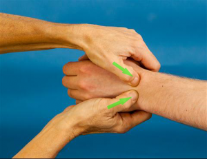 Another less commonly injured ligament of the wrist is the lunotriquetral (LT). The mechanism of injury of the SL and LT ligaments is different. The SL is potentially injured in wrist flexion, radial deviation, and supination, whereas the LT occurs in wrist extension, radial deviation, and pronation. The test for the LT ligament is the LT shearing sign, AKA Reagan test. It is performed in sitting with the elbow flexed and forearm in neutral. The clinician places one thumb on the lunate, one thumb on triquetrum, and a shear force is applied in the anterior/posterior plane by alternating pressure between the 2 contacts. A positive test is pain, laxity, or crepitis. The statistical data reveals this test to be a modest tool for assessment of the LT ligament: Sensitivity = 66 – 95%; Specificity = 64 – 87%.
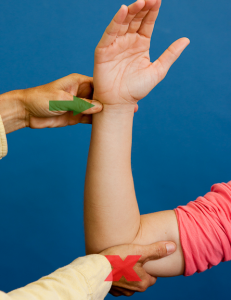 Finally, the Fovea sign assesses disruptions of the distal radio-ulnar ligament and ulno-triquetral ligament. To perform this test, the elbow is placed in 90° of flexion, forearm and wrist in neutral and the clinician presses his/her thumb into the soft spot between the pisiform and ulnar styloid. Remember, you are assessing the distal radio-ulnar ligament and ulno-triquetral ligament, but if you go too proximal with your palpation, you could be on the triangular fibrocartilage. Thus, you want to make sure your palpation is precise. A positive test is defined as “exquisite pain.” Despite the potential for palpation error, sensitivity and specificity are excellent for this test: Sensitivity = 95.2%; Specificity = 86.5%; (+) LR = 1.69-7.1; (-) LR = 0.06-0.56
In summary, the assessment of wrist stability is important. The wrist has a critical relationship with the function of the hand. Wrist stability is needed for grip strength and fine motor dexterity. Assessing the wrist properly will increase the likelihood of an accurate diagnosis and an appropriate intervention. For videos of all of these tests and more cutting edge orthopedic information in iOrtho+ Premium Mobile App, please visit https://iortho.xyz/
- Dutton M, 2004 Orthopedic examination, evaluation & intervention, New York, McGraw Hill
- Foreman TA, Foreman SK, Rose NE, 2005 Clinical approach to diagnosing wrist pain. American Family Physician. 72(9):1753-1758.
- Gulick, DT, 2018 OrthoNotes, 4th FA Davis Publishing, Philadelphia
- Gulick DT, 2020 iOrtho+ Mobile App. DTG Enterprises LLC
- Lan LB, 1995 The scaphoid shift test. The Journal of Hand Surgery. 1993;18A(2):366-368
- LaStayo P, Howell J. Clinical provocative tests used in evaluating wrist pain: a descriptive study. Journal Hand Therapy. 8:10-17
- Magee D, 2008. Orthopedic Physical Assessment. 5th ed. Philadelphia, PA: WB Saunders Company
- Marx RG, Bombardier C, Wright JC, 1999 What do we know about the reliability & validity of physical examination tests used to examine the upper extremity? Journal of Hand Surgery. 24A:185-193
- Powell JM, Lloyd GJ, Rintoul RF, 1988 New clinical test for fracture of the scaphoid. Canadian Journal Surgery. 31:237-238
- Shin AY, Battaglia MJ, Bishop AT, 2000 Lunotriquetral instability: diagnosis and treatment, J Am Acad Orthop Surg 8:170-179
- Taleisnik J, 1988 Carpal instability, Journal Bone Joint Surgery American. 70:1262-1268
- Tay SC, Tomita K, Berger RA, 2007 The ulnar fovea sign for defining ulnar wrist pain: an analysis of sensitivity and specificity. Journal Hand Surgery American. 32(4):438-44
- Valadic AL, Jobe CM, Pink MM et al, 2000 Anatomy of provocative tests for impingement syndrome of the shoulder, Journal Shoulder Elbow Surgery. 9:36-46
- Waeckerle JF, 1987 A prospective study identifying the sensitivity of radiographic findings & the efficacy of clinical findings in carpal navicular fractures. Annals Emergency Medicine. 16:733-737.
- Watson HK, Ashmead D, Makhlouf MV, 1988 Examination of the scaphoid, Journal Hand Surgery American. 13:657-660
- Watson HK, Ballet FL, 1984 The SLAC wrist: scapulolunate advanced collapse pattern of degenerative arthritis, Journal Hand Surgery American. 9:358-365
- Young D, Papp S, Giachino A, 2010 Physical examination of the wrist. Hand Clinics. 26(1):21-36
- Young DK, Giachino A, 2009 Clinical examination of scaphoid fractures. Physician & Sports Medicine. 37(1):97-105